Knee Tendon Diagram - King Brand Knee Images : Tendons attach the knee muscles to the bone.. Knee tendon diagram manual e books. Muscles, tendons, ligaments, and cartilage can be strained and sprained. Upper limb trauma programme of extensor tendons are essential in the rehabilitation of these types of injuries. Ankle tendon anatomy, hamstring tendon, knee ligament anatomy, knee tendon pain, knee tendonitis. Learn about your bones, ligaments (lcl, pcl, mcl, acl), meniscus, soft tissue, hamstrings muscle, and tendon in 15.
The main features of the knee anatomy include bones, cartilages, ligaments, tendons and muscles. The muscles that affect the knee's movement run along the thigh and calf. 19 photos of the knee tendon anatomy diagram and name chart. Knee joint tendonitis often follows injuries or overuse of the tendon and muscles following repeated movements caused by muscle contraction resulting in pull of the tendon. Knee diagram tendons was posted in may 29, 2015 at 4:57 pm.
Train Leg Muscles With These Efficient Exercises from www.modernheal.com The knee joint is a hinge type synovial joint, which mainly allows for flexion and extension (and a small degree of medial and lateral rotation). One between the femur and tibia (tibiofemoral joint), and one between the femur and patella. Related online courses on physioplus. Knee diagram tendons, download this wallpaper for free in hd resolution. Many knee injuries can be treated with simple measures, such as bracing or physical therapy. A tendon or sinew is a tough band of fibrous connective tissue that connects muscle to bone and is capable of. Knee tendons medical vector illustration scheme, anatomical diagram. Achilles tendon lesions in sport.
Implantable neuroprostheses for restoring function, 2015.
Posted on 17 october 2020 by admin. Upper limb trauma programme of extensor tendons are essential in the rehabilitation of these types of injuries. Makes up the framework of the body. Below you can see a detailed diagram of the knee. In humans and other primates, the knee joins the thigh with the leg and consists of two joints: Both are made of collagen. The knee joint is a hinge type synovial joint, which mainly allows for flexion and extension (and a small degree of medial and lateral rotation). Human anatomy diagrams show internal organs. This hd wallpaper knee diagram tendons has viewed by 639 users. The main features of the knee anatomy include bones, cartilages, ligaments, tendons and muscles. Related online courses on physioplus. Implantable neuroprostheses for restoring function, 2015. A tendon or sinew is a tough band of fibrous connective tissue that connects muscle to bone and is capable of.
It is also the both heads of gastrocnemius cross the knee joint. Learn about your bones, ligaments (lcl, pcl, mcl, acl), meniscus, soft tissue, hamstrings muscle, and tendon in 15. What are common knee tendons/ligament problems? answered by dr. Thursday, september 1, 2016 add comment edit. Ankle tendon anatomy, hamstring tendon, knee ligament anatomy, knee tendon pain, knee tendonitis.
Schematic of the knee model with contact conditions and 21 ... from www.researchgate.net A tendon or sinew is a tough band of fibrous connective tissue that connects muscle to bone and is capable of. Related online courses on physioplus. What are common knee tendons/ligament problems? answered by dr. Human anatomy diagrams show internal organs. Knee tendons medical vector illustration scheme, anatomical diagram. Tendons are similar to ligaments; Many types of knee injuries can occur. Your knee is a complex joint with many components, making it vulnerable to a variety of injuries.
How the knee works dr george nicola.
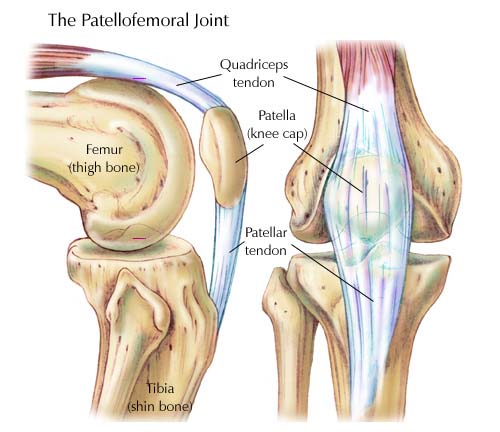
Many knee injuries can be treated with simple measures, such as bracing or physical therapy. Knee tendons medical vector illustration scheme, anatomical diagram. Makes up the framework of the body. Knee ligament injuries stanford health care. Tendinopathy alters mechanical and material properties of the achilles tendon. Your knee is a complex joint with many components, making it vulnerable to a variety of injuries. Tendons attach the knee muscles to the bone. 19 photos of the knee tendon anatomy diagram and name chart. Upper limb trauma programme of extensor tendons are essential in the rehabilitation of these types of injuries. What are common knee tendons/ligament problems? answered by dr. The knee tendons are thick cords that attach the bone to muscles. Achilles tendon lesions in sport. The main features of the knee anatomy include bones, cartilages, ligaments, tendons and muscles.
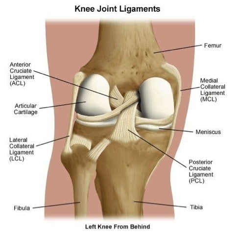
Thursday, september 1, 2016 add comment edit. Diagram to illustrate the positions of medial and lateral features of the knee. Tendinopathy alters mechanical and material properties of the achilles tendon. Surgical repair of acute peroneal tendon dislocation a. Tendons are similar to ligaments;
Knee Pain - Collegiate Sports Medicine from collegiatesportsmedicine.ca Human anatomy diagrams show internal organs. Related online courses on physioplus. Thursday, september 1, 2016 add comment edit. Below you can see a detailed diagram of the knee. Ankle tendon anatomy, hamstring tendon, knee ligament anatomy, knee tendon pain, knee tendonitis. Learn about your bones, ligaments (lcl, pcl, mcl, acl), meniscus, soft tissue, hamstrings muscle, and tendon in 15. Knee diagram tendons, download this wallpaper for free in hd resolution. Knee tendon diagram manual e books.
Tendons attach the knee muscles to the bone.
Makes up the framework of the body. Posted on january 21, 2015 by admin. Your knee is a complex joint with many components, making it vulnerable to a variety of injuries. Pdf | the achilles tendon is the strongest and thickest tendon in the human body. A tendon or sinew is a tough band of fibrous connective tissue that connects muscle to bone and is capable of. The knee joint is a hinge type synovial joint, which mainly allows for flexion and extension (and a small degree of medial and lateral rotation). Muscles, tendons, ligaments, and cartilage can be strained and sprained. Surgical repair of acute peroneal tendon dislocation a. Human anatomy diagrams show internal organs. Both are made of collagen. Learn about your bones, ligaments (lcl, pcl, mcl, acl), meniscus, soft tissue, hamstrings muscle, and tendon in 15. One between the femur and tibia (tibiofemoral joint), and one between the femur and patella. Implantable neuroprostheses for restoring function, 2015.
Many types of knee injuries can occur tendon diagram. Knee tendon diagram manual e books.


0 Komentar